Verified institutional research accounts only. Supplied for in vitro laboratory research and preclinical studies.
Add to Cart
First-time checkout is released after institutional account review.
| Intra-Assay CV: | <10% | ||||||||||||||||||||||||||||||||||||||
| Inter-Assay CV: | <15% | ||||||||||||||||||||||||||||||||||||||
| Sensitivity: | 0.08 ng/ml | ||||||||||||||||||||||||||||||||||||||
| Specificity: |
| ||||||||||||||||||||||||||||||||||||||
| Linear Range: | 0.08 - 0.7 ng/ml | ||||||||||||||||||||||||||||||||||||||
| Sample Extraction: | recommended | ||||||||||||||||||||||||||||||||||||||
| Standard Curve: | 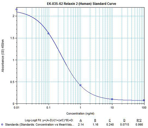 |
Contact us for technical specifications on this product.