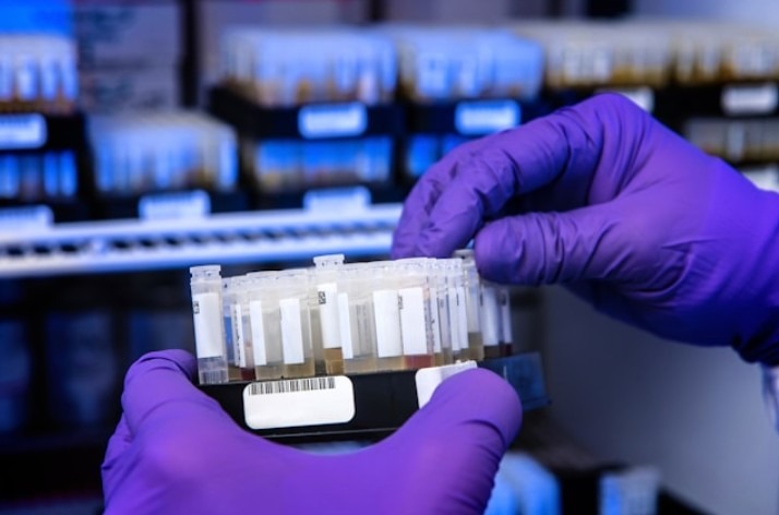
Polyclonal Antibodies
What Are Polyclonal Antibodies?
Polyclonal antibodies are a type of antibody produced by multiple B-cell clones in response to an antigen. Unlike monoclonal antibodies, which are derived from a single immune cell, polyclonal antibodies recognize multiple epitopes on the same antigen. This multi-epitope binding enhances sensitivity, making them ideal for detecting low-abundance targets and improving assay performance. Phoenix Pharmaceuticals specializes in producing high-affinity polyclonal antibodies to support a wide range of scientific disciplines.
Applications of Polyclonal Antibodies in Research
Researchers across various fields rely on polyclonal antibodies for numerous applications, including:
- Immunohistochemistry (IHC): Detecting specific proteins in tissue samples for disease research.
- Western Blotting: Identifying target proteins in complex biological samples.
- Enzyme-Linked Immunosorbent Assay (ELISA): Quantifying protein levels in diagnostic and experimental assays.
- Flow Cytometry: Characterizing cell populations based on protein expression.
Due to their high sensitivity and broad epitope recognition, polyclonal antibodies are particularly valuable in detecting conformational changes and post-translational modifications in proteins.

The Challenges in Producing High-Quality Polyclonal Antibodies
Producing high-quality polyclonal antibodies requires precision, expertise, and stringent quality control. At Phoenix Pharmaceuticals, we are committed to overcoming the challenges associated with antibody production to ensure that researchers receive antibodies with superior specificity, sensitivity, and reproducibility.
Ensuring High Specificity and Affinity
One of the primary challenges in polyclonal antibody production is achieving high specificity while maintaining strong binding affinity. Because polyclonal antibodies recognize multiple epitopes on an antigen, there is a risk of cross-reactivity with unintended targets. To mitigate this, we carefully design immunogens, optimize antigen purity, and use rigorous screening processes to select antibodies with the highest specificity.
Maintaining Consistency Across Batches
Since polyclonal antibodies are derived from multiple B-cell clones within an immunized host, variability between production batches can be a concern. To ensure consistency, we standardize immunization protocols, employ strict purification techniques, and conduct extensive validation testing on each batch. This guarantees that researchers receive reproducible results across experiments.
Optimizing Purification and Yield
Extracting and purifying polyclonal antibodies from serum involves complex fractionation and filtration processes. Achieving a high yield without compromising purity requires advanced purification techniques such as affinity chromatography. At Phoenix Pharmaceuticals, we use state-of-the-art purification methods to remove unwanted serum proteins and isolate high-affinity antibodies with minimal contaminants.
Ensuring Stability and Longevity
Polyclonal antibodies must remain stable during storage and transportation to maintain their efficacy. Factors such as temperature fluctuations, pH variations, and oxidation can degrade antibody integrity. To address this, we provide antibodies in optimized storage buffers and conduct stability testing to ensure long-term viability.
By addressing these challenges with rigorous quality control, advanced production techniques, and continuous optimization, Phoenix Pharmaceuticals delivers polyclonal antibodies that researchers can trust for their most demanding applications.
Who Uses Phoenix Pharmaceuticals’ Polyclonal Antibodies?
Our polyclonal antibodies are trusted by:
- Academic researchers studying molecular biology, neuroscience, immunology, and more.
- Pharmaceutical and biotechnology companies conducting drug discovery and biomarker validation.
- Diagnostic laboratories developing new assays for disease detection and monitoring.
- Government and clinical research institutions focused on advancing biomedical science.
Phoenix Pharmaceuticals understands that high-quality reagents are essential for reliable research outcomes. Our polyclonal antibodies are meticulously developed and validated to ensure specificity, consistency, and reproducibility. If it’s off-the-shelf solutions or custom antibody production, we are here to support your research needs.
Explore our collection of polyclonal antibodies today and take your research to the next level with Phoenix Pharmaceuticals.
Highly-specific polyclonal antibodies raised in-house.
*Products listed in alphabetical order. If you are looking for something specific, type it into the Search Bar at the top of the website.
