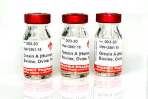
Catalog # |
Size |
Price |
|
---|---|---|---|
030-30 | 100 µg | $423 |
Asn-Pro-Cys-Asn-Ala-Glu-Phe-Gln-Asn-Phe-Cys-Ile-His-Gly-Glu-Cys-Lys-Tyr-Ile-Glu-His-Leu-Glu-Ala-Val-Thr-Cys-Lys-Cys-Gln-Gln-Glu-Tyr-Phe-Gly-Glu-Arg-Cys-Gly-Glu-Lys
|
Disulfide bridge(s): C1-C3, C2-C4 & C5-C6
|
4794.38 | |
| ≥ 95% |
| Exhibits correct molecular weight |
| Soluble in water |
|
Up to 6 months in lyophilized form at 0-5°C. For best results, rehydrate just before use. After rehydration, keep solution at +4°C for up to 5 days or freeze at -20°C for up to 3 months. Aliquot before freezing to avoid repeated freeze-thaw cycles. |
| White powder |
| Each vial contains 100 µg of NET peptide. |
The EGFR ligand amphiregulin (AREG) has been implicated as an important autocrine growth factor in several epithelial malignancies and in psoriasis, a hyperproliferative skin disorder. To characterize the mechanisms by which AREG regulates autocrine epithelial cell growth, we transduced human keratinocytes (KCs) with lentiviral constructs expressing tetracycline (TET)-inducible small hairpin RNA (shRNA). TET-induced expression of AREG shRNA markedly reduced autocrine extracellular signal-regulated kinase phosphorylation, strongly inhibited autocrine KC growth with an efficiency similar to metalloproteinase and EGFR inhibitors, and induced several markers of KC differentiation, including keratins 1 and 10. Addition of various concentrations of exogenous EGFR ligands to KC cultures reversed the growth inhibition in response to AREG-blocking antibodies but not to shRNA-mediated AREG knockdown. Lentivirus-mediated expression of the full-length AREG transmembrane (TM) precursor, but not of the AREG extracellular domain, markedly reversed the shRNA-mediated growth inhibition and morphological changes, and strongly reduced the induction of multiple markers of KC differentiation. Taken together, our data show that autocrine human KC growth is highly dependent on the AREG TM precursor protein and strongly suggest a previously unreported function of the metalloproteinase-processed carboxy (C)-terminal domain of AREG.
Stoll SW, Johnson JL, Li Y, Rittié L, Elder JT. Amphiregulin carboxy-terminal domain is required for autocrine keratinocyte growth. J Invest Dermatol. 2010;130(8):2031-40.
Amphiregulin (AREG) is an important regulator of cellular growth in keratinocytes, carcinomas, and hyperproliferative epidermal disorders, including psoriasis. Stoll and colleagues present data suggesting that the cytoplasmic carboxy-terminal domain of amphiregulin plays an important role in regulating autocrine keratinocyte growth through the epidermal growth factor receptor. These observations raise novel and interesting biological questions regarding the function of the cytoplasmic C-terminal region of AREG.
Seykora JT. Grabbing amphiregulin by the tail to better understand keratinocyte growth. J Invest Dermatol. 2010;130(8):1966-8.
The regulation of amphiregulin, an epidermal growth factor (EGF) family member, and its effect on vascular smooth muscle cells (VSMC) were examined. Amphiregulin mRNA was upregulated by amphiregulin itself as well as alpha-thrombin. Amphiregulin caused an approximate 3-fold increase in DNA synthesis. Its effect on growth was compared with those of other mitogens, and was found to be approximately 3.5-, 2.4-, and 1.0-fold greater than those of endothelin-I (ET-I), alpha-thrombin, and platelet-derived growth factor-AB (PDGF-AB), respectively. As evidenced by Western blot analysis, amphiregulin stimulated the phosphorylation of p42/p44-mitogen-activated protein kinase (MAPK), p38-MAPK, c-Jun NH2-terminal protein kinase (JNK), and Akt/protein kinase B (PKB), respectively. By statistical analysis, the amphiregulin-induced growth effect was significantly decreased by the MAP kinase/ extracellular regulated kinase kinase-1 (MEK-1) inhibitor PD98059, p38-MAPK inhibitor SB203580, and phosphatidylinositol 3-kinase (PI-3 kinase) inhibitor wortmannin, respectively, but was not decreased by JNK inhibitor SP600125. These results suggest that amphiregulin is the most potent mitogen of the mitogens tested, and its growth effect is mediated at least in part through the p42/p44-MAPK, p38-MAPK, and PI-3 kinase-Akt/PKB pathways in VSMC.
Kato M, Inazu T, Kawai Y, et al. Amphiregulin is a potent mitogen for the vascular smooth muscle cell line, A7r5. Biochem Biophys Res Commun. 2003;301(4):1109-15.
Several members of the epidermal growth factor (EGF) family of growth factors that contain EGF-like units at their carboxy portion have been isolated and characterized. Schwannoma-derived growth factor (SDGF) and amphiregulin (AR) are members of this family. SDGF has high sequence homology to AR, and is known to be not only a potent mitogen for astrocytes and fibroblasts but also a neurotrophic factor. We previously confirmed that the synthetic EGF-like peptides SDGF(38-80) and AR(44-84), corresponding to the EGF-like domain of mouse SDGF and human AR, respectively, formed similar disulfide bond patterns to that of EGF. In the present study, we further investigated the biological actions of these two EGF-like peptides on several cultured cell lines. We found that SDGF(38-80) and AR(44-84) have weak mitogenic activity in NIH/3T3 cells and weak binding affinity to the EGF receptor on the surface of A431 cells compared with EGF. However, SDGF(38-80) and EGF induced short neurite outgrowth in PC12 h cells, a subclone of PC12 cells, at 100 nM. In addition, a significant increase in acetylcholinesterase (AChE) activity induced by SDGF(38-80) was observed at a concentration similar range to that of EGF, which is known as a differentiation marker of these cells. The effect of AR(44-84) in PC12 h cells was weaker than those of SDGF(38-80) and EGF, but the AChE activity was significantly increased by the addition of 100 nM AR(44-84), which did not stimulate NIH/3T3 cell growth. These results also suggest that SDGF(38-80) and AR(44-84) may be effective for neuronal differentiation rather than proliferation.
Takenouchi T, Kadosaka M, Shin SY, Munekata E. Biological actions of the epidermal growth factors-like domain peptides of mouse schwannoma-derived growth factor and human amphiregulin. J Pept Res. 1999;53(2):120-5.
Biosynthesis and processing of amphiregulin (AR) have been investigated in human colorectal (HCA-7, Caco-2) and mammary (MCF-7) cancer cell lines, as well as in Madin-Darby canine kidney cells stably expressing various human AR precursor (pro-AR) forms. Both cells expressing endogenous and transfected AR produce multiple cellular and soluble forms of AR with an N-glycosylated 50-kDa pro-AR form being predominant. Our results demonstrate that sequential proteolytic cleavage within the ectodomain of the 50-kDa pro-AR form leads to release of a predominant N-glycosylated 43-kDa soluble AR, as well as the appearance of other cellular and soluble AR forms. Cell surface biotinylation studies using a C-terminal epitope-tagged pro-AR indicate that all cell surface forms are membrane-anchored and support that AR is released by ectodomain cleavage of pro-AR at the plasma membrane. We also show that pro-AR ectodomain cleavage is a regulated process, which can be stimulated by phorbol 12-myristate 13-acetate and inhibited by the metalloprotease inhibitor, batimastat. In addition, we provide evidence that high molecular mass AR forms may retain the full-length N-terminal pro-region, which may influence the biological activities of these forms.
Brown CL, Meise KS, Plowman GD, Coffey RJ, Dempsey PJ. Cell surface ectodomain cleavage of human amphiregulin precursor is sensitive to a metalloprotease inhibitor. Release of a predominant N-glycosylated 43-kDa soluble form. J Biol Chem. 1998;273(27):17258-68.
No References
Catalog# | Product | Size | Price | Buy Now |
---|
Social Network Confirmation